Clipart microscope parts labeled WikiClipArt
Parts of the Microscope (Labeled Diagrams) By Editorial Board December 14, 2022 The microscope is one of the must-have laboratory tools because of its ability to observe minute objects, usually living organisms that cannot be seen by the naked eyes. It is categorized into two: simple and compound microscopes.

Parts of a microscope with functions and labeled diagram
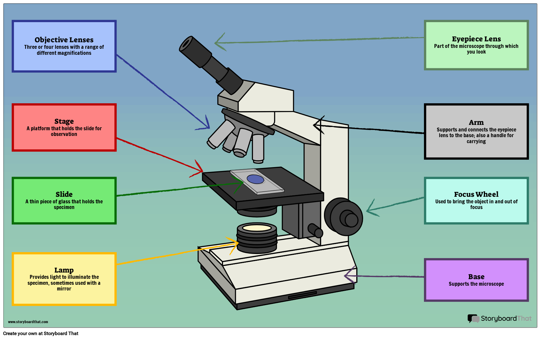
Drag and drop the text labels onto the microscope diagram.

301 Moved Permanently
Fluorescence microscopes: These use fluorescent dyes to highlight specific structures or molecules in a sample and are commonly used in biological research. X-ray microscopes: These use X-rays to produce images of the internal structure of samples and are often used to study materials and biological specimens.
Parts Of Microscope
A labeled diagram of microscope parts furnishes comprehensive information regarding their composition and spatial arrangement within the microscope, enabling researchers to comprehend their function effectively. In this comprehensive article, we will delve into the intricate parts of the microscope, exploring their functions in detail.
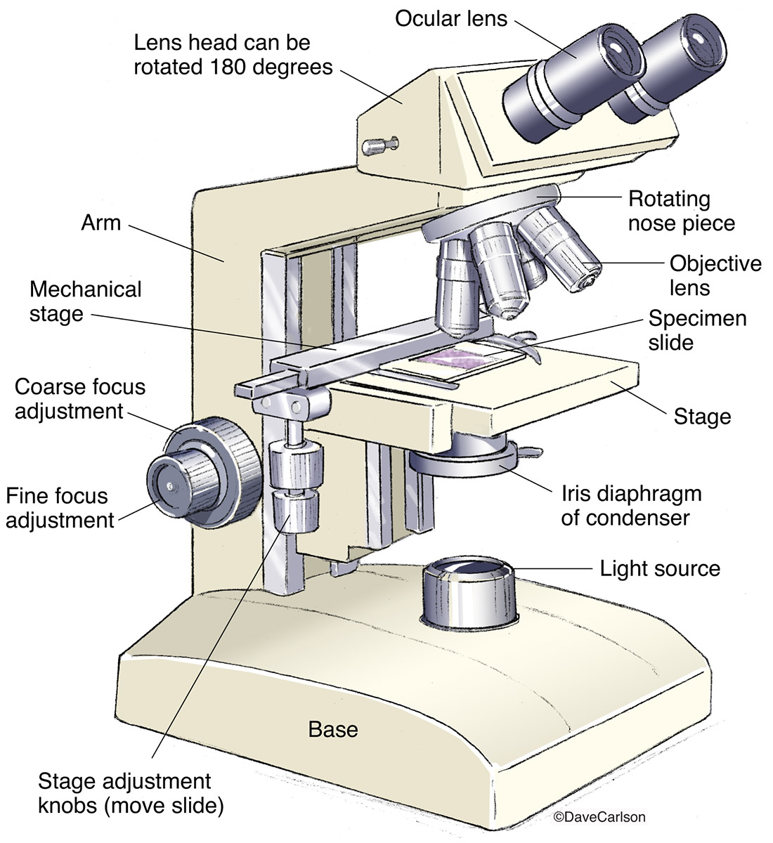
Compound Microscope Carlson Stock Art
The web page titled "Parts of a Microscope with Labeled Diagram and Functions" has the following key takeaways: 🔍 The microscope is an essential tool for scientists, researchers, and medical professionals. 🧬 The main function of a microscope is to provide a magnified view of small objects or organisms, such as bacteria, cells, or tissues.

5 Types of Microscopes with Definitions, Principle, Uses, Labeled Diagrams
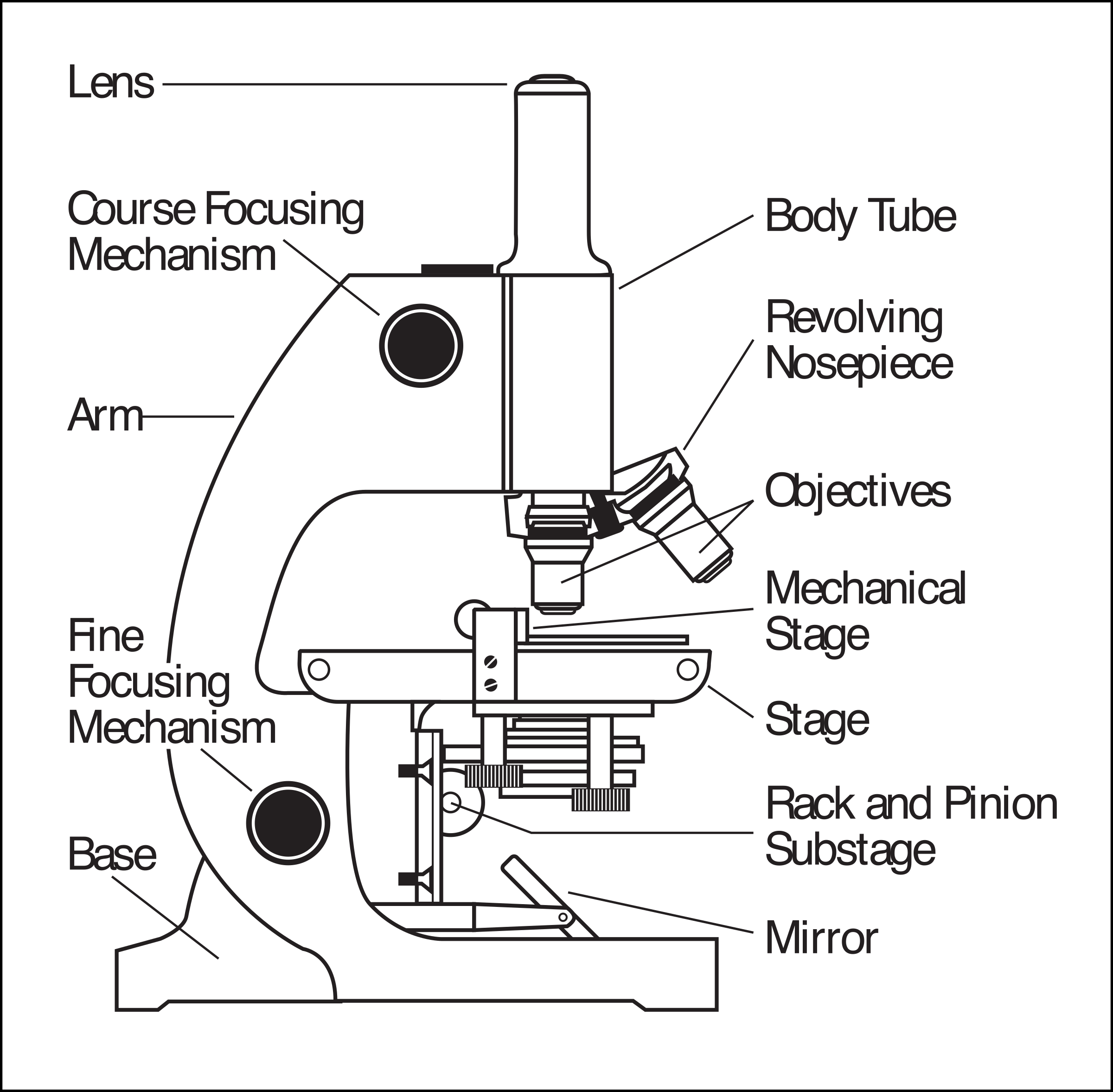
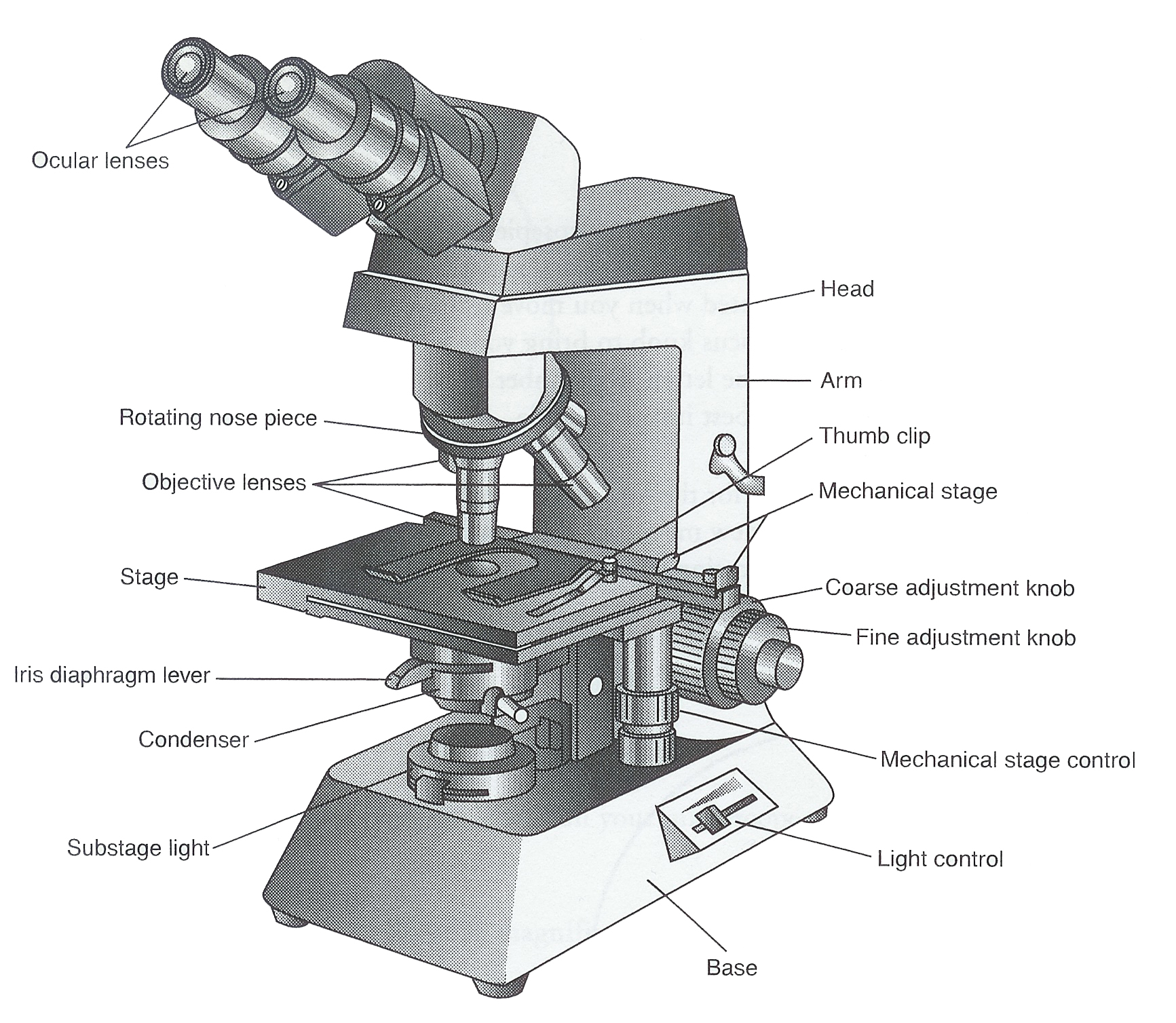
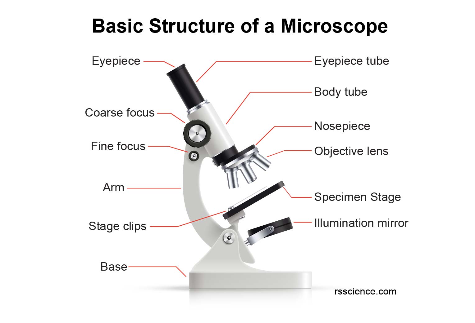
Figure: Diagram of parts of a microscope. There are three structural parts of the microscope i.e. head, arm, and base. Head - The head is a cylindrical metallic tube that holds the eyepiece lens at one end and connects to the nose piece at other end. It is also called a body tube or eyepiece tube.
Monday September 25 Parts of a Compound Light Microscope
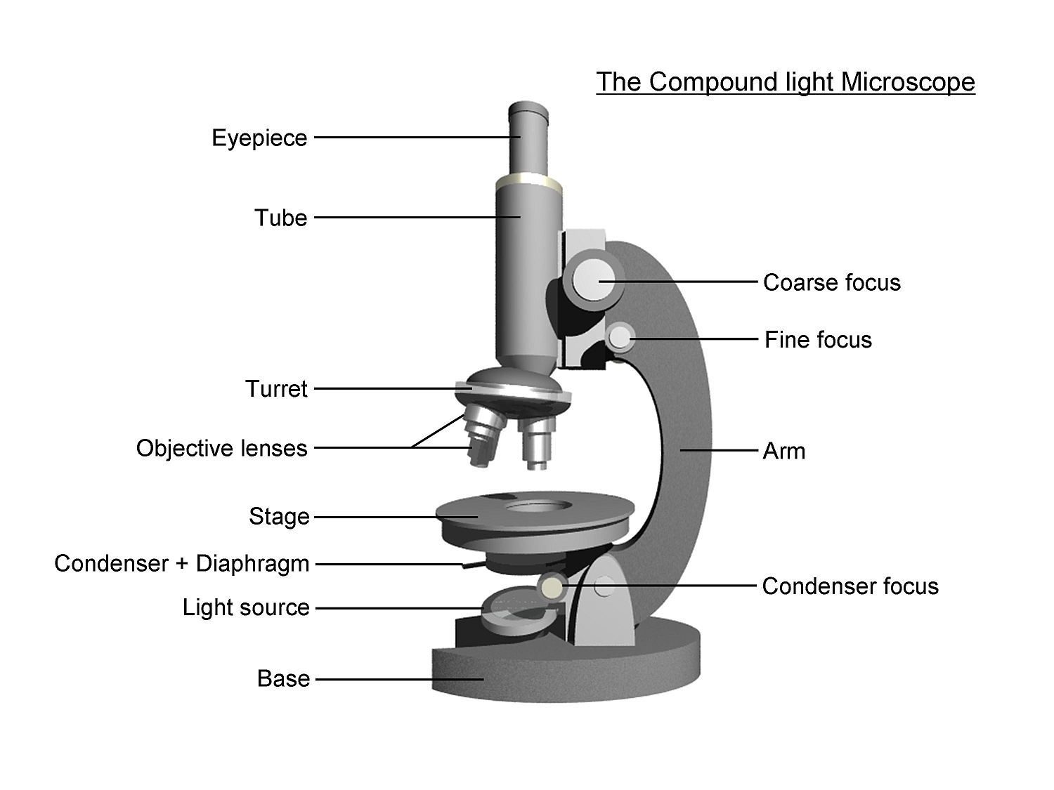
The optical microscope often referred to as the light microscope, is a type of microscope that uses visible light and a system of lenses to magnify images of small subjects. There are two basic types of optical microscopes: Simple microscopes. Compound microscopes. The term "compound" in compound microscopes refers to the microscope having.
Ag Biology Unit 2
A microscope is an optical instrument used to magnify an image of a tiny object;. Diagram. Also see : Labeling the parts of the Microscope. Image source: slidesharecdn.com. Simple microscope Parts and Functions. A simple microscope's parts have two classifications: the mechanical part and the optical parts.
Cells and Microscopes
This activity has been designed for use in homes and schools. Each microscope layout (both blank and the version with answers) are available as PDF downloads. You can view a more in-depth review of each part of the microscope here. Download the Label the Parts of the Microscope PDF printable version here.
1.5 Microscopy Biology LibreTexts
These diagrams clearly explain the functioning of the microscopes along with their respective parts. Man's curiosity has led to great inventions. The microscope is one of them. Probably, it all started when man, realized that a piece of crystal could magnify images because it is thicker in the center than in the edges.
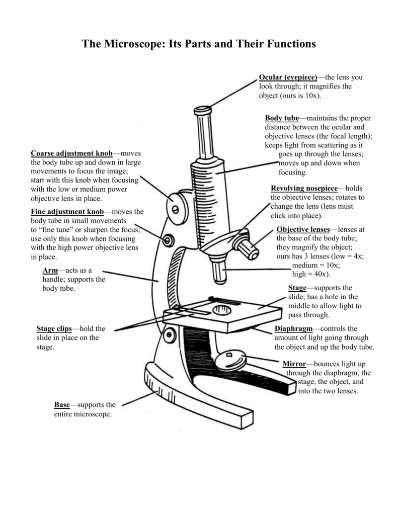
The Microscope Its Parts and Their Functions
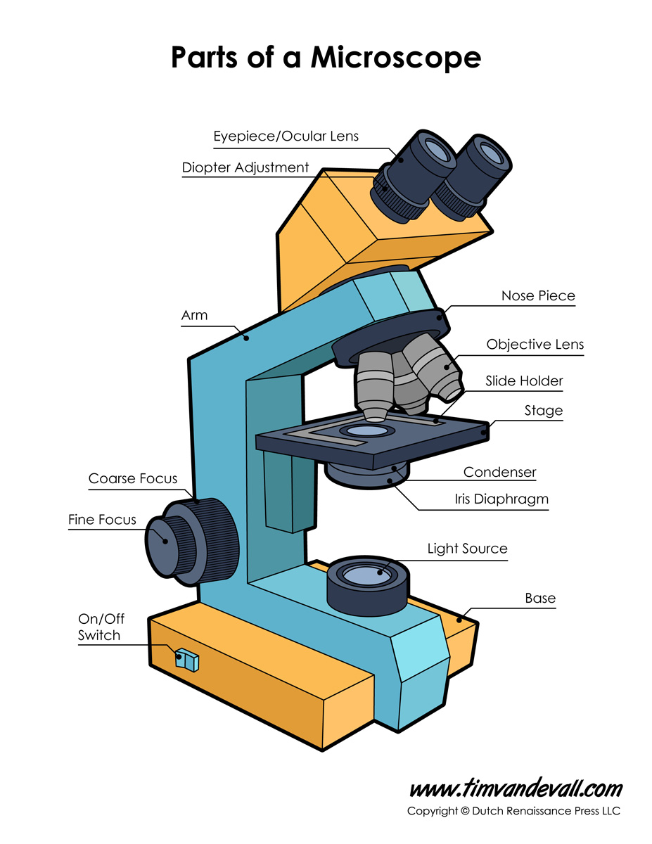
Included in this list are the following elements: 1 . Eyepiece : It is also known as the ocular, is the portion of the microscope through which you view the specimen under examination. 2. Tube for the eyepiece: A holder for an eyepiece is precisely what it sounds like. The eyepiece is located above the objective lens.
16 Parts of a Compound Microscope Diagrams and Video Microscope Clarity
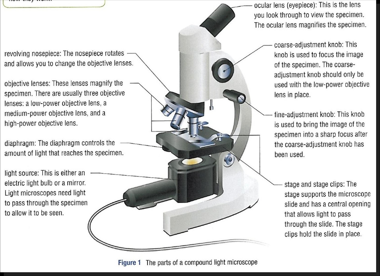
Ocular Lens (eye-piece) Ocular lens of a microscope. It is located at the top of the microscope, and the ocular lens or eyepiece lens is used to look through the specimen. It also magnifies the image formed by the objective lens, usually ten times (10x) or 15 times (15x). Usually, a microscope has an eyepiece of 10x magnification power.

Parts of a Compound Microscope Labeled (with diagrams) Medical
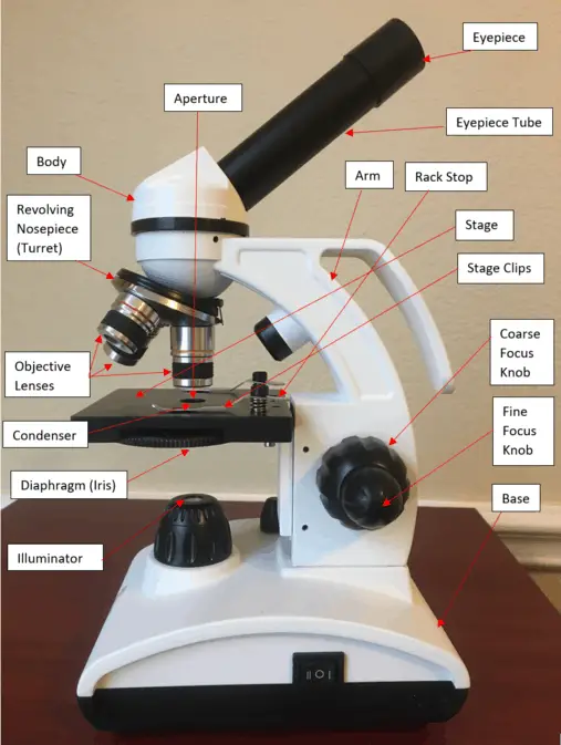
1. Eyepiece 2. Body tube/Head 3. Turret/Nose piece 4. Objective lenses 5. Knobs (fine and coarse) 6. Stage and stage clips 7. Aperture 9. Condenser 10. Condenser focus knob 11. Iris diaphragm 12. Diopter adjustment 13. Arm 14. Specimen/slide 15. Stage control/stage height adjustment 16. On and off switch 17. Base
What is a Microscope? Function and Magnification Rs' Science
This article covers An overview of microscopes What is a "compound microscope"? Labeled diagram of a compound microscope Major structural parts of a compound microscope Optical components of a compound microscope Eyepiece Eyepiece tube Objective lenses Nosepiece Specimen stage Coarse and fine focus knobs Rack stop Illuminator Condenser
Parts of a Microscope Labeling Activity
Figure: Labeled Diagram of a Light Microscope. Types of light microscopes (optical microscope) With the evolved field of Microbiology, the microscopes. used to view specimens are both simple and compound light microscopes, all using lenses. The difference is simple light microscopes use a single lens for magnification while compound lenses use.

Microscope diagram Tom Butler Technical Drawing and Illustration
The working principle of a simple microscope is that when a lens is held close to the eye, a virtual, magnified and erect image of a specimen is formed at the least possible distance from which a human eye can discern objects clearly. Magnification formula The magnification power of a simple microscope is expressed as: M = 1 + D/F Where